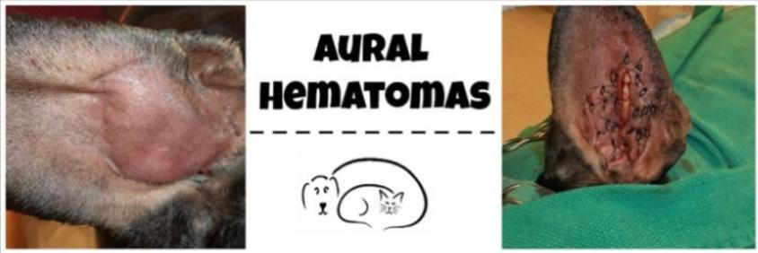

Recurrence is common with needle drainage alone, even if bandages are applied, but treatment with systemic or intralesional steroids reduces recurrence. Nonsurgical treatment involves needle drainage and flushing of the hematoma cavity. Drains are removed within 1 week, but bandaging may be required for 2 additional weeks.
AURAL HEMATOMA SURGERY SKIN
Apposition of skin to cartilage is maintained by placement of sutures between skin and cartilage, application of a compressive bandage, or both. Surgical options include placement of an active or passive drain or fenestration on the concave skin of the pinna, using either a single long incision or multiple small incisions, to empty the pocket and prevent recurrence of fluid accumulation. Drainage should be performed expeditiously to prevent contracture, fibrosis, and subsequent deformity.

Therapeutic goals include removal of contents, maintenance of apposition between skin and cartilage, and appropriate treatment for the source of head shaking or scratching. Related Article: Diagnosis & Management of Otitis TreatmentĪural hematomas can be treated surgically or nonsurgically. Affected animals should undergo thorough dermatologic and otoscopic examinations, and any underlying disease should be treated to facilitate resolution and prevent recurrence. If untreated, granulation tissue replaces aural hematomas subsequent contraction and fibrosis of this tissue can result in pinnal deformity and, in cats, obstruction of the external acoustic opening.Īural hematomas can be caused by direct damage (eg, bite wounds, vehicular trauma) but are more common from head shaking and ear scratching associated with otitis externa or atopy. Hemorrhage from the penetrating vessels continues, ultimately forming an out-pouching (ie, aural hematoma). Normally, the concave surface of pinna cartilage is firmly attached to the skin, but trauma may create dead space between the cartilage and skin that fills with blood. Branches of the caudal auricular artery passing from the convex to the concave surface of the pinna through tiny channels within the cartilage provide blood to the auricular cartilage.


 0 kommentar(er)
